
If your doctor has recommended an EEG, you may be wondering what the test actually does, whether it hurts, and what the results may tell you.
An EEG test, short for electroencephalogram, records the electrical activity of your brain through small electrodes placed on your scalp. It is used most often when a doctor is investigating seizures, epilepsy, staring spells, unexplained blackouts, or unusual changes in awareness.
The electrodes only record signals. They do not send electricity into your brain, and a routine EEG is generally painless.
Medically guided by Dr. Siddharth Kharkar
Trusted neurological guidance that turns confusing symptoms and test results into clearer next steps. The aim is not simply to perform an EEG, but to understand whether your symptoms, clinical history, examination, and test findings fit together.
Quick Answer: What Is an EEG Test?
An EEG is a neurological test that records the brain’s natural electrical activity.
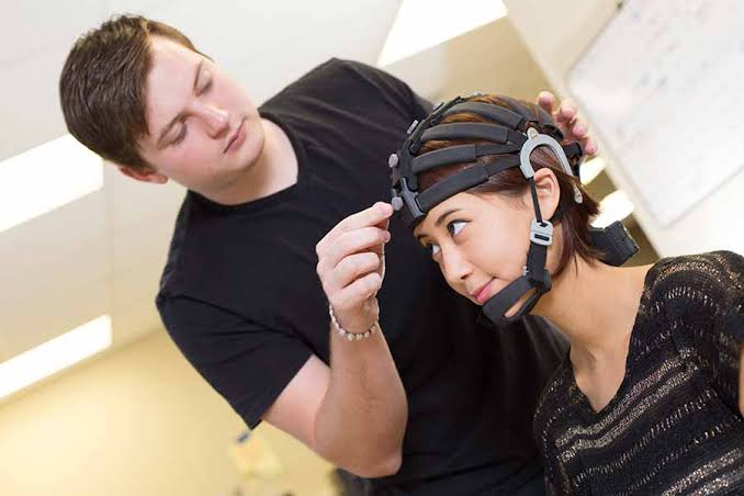
A technician places small electrodes on your scalp while you sit or lie comfortably. During the recording, you may be asked to close your eyes, breathe deeply, or look at flashing lights. These steps can make certain brain-wave patterns easier to detect.
EEG is particularly useful when seizures or epilepsy are suspected. However, it is only one part of a neurological diagnosis. A normal EEG does not automatically mean that a seizure did not occur.
At a Glance
- What does EEG measure? Electrical activity produced by the brain.
- Does it hurt? Usually no.
- Does electricity enter the brain? The electrodes only record activity.
- Why is it ordered? Most often for seizures, suspected epilepsy, unexplained loss of awareness, or related neurological concerns.
- How long does it take? A routine recording is commonly around 20–40 minutes, although preparation makes the complete appointment longer.
- Can a normal EEG rule out epilepsy?
- Can EEG diagnose a stroke by itself? Stroke and TIA evaluation usually depends much more on clinical assessment and brain or vascular imaging.
What Is an EEG and What Does It Measure?
Your brain cells communicate through electrical signals. An electroencephalogram detects patterns created by that electrical activity.
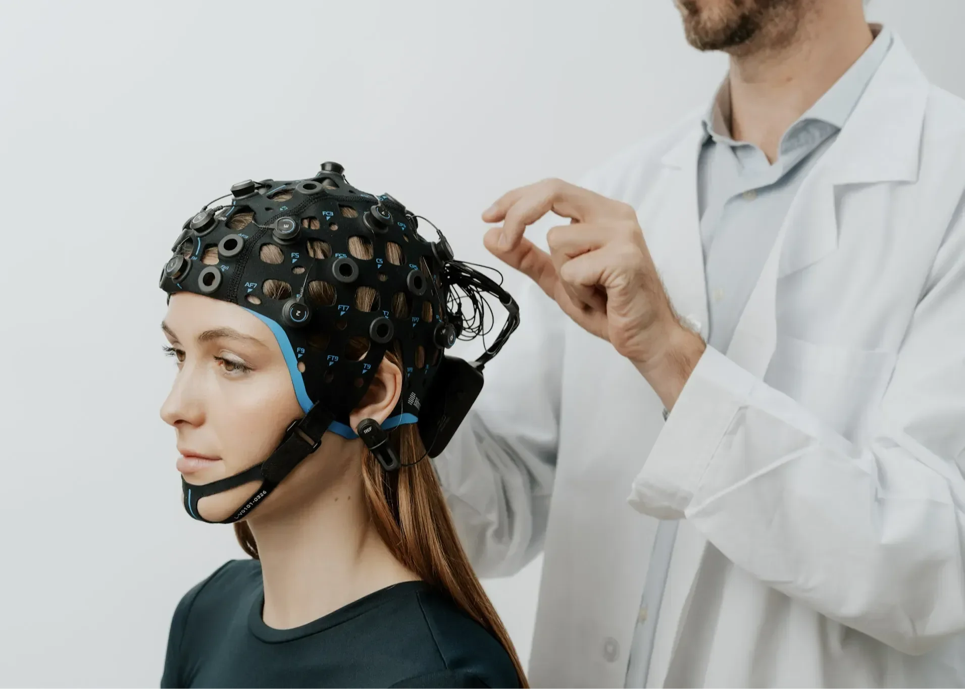
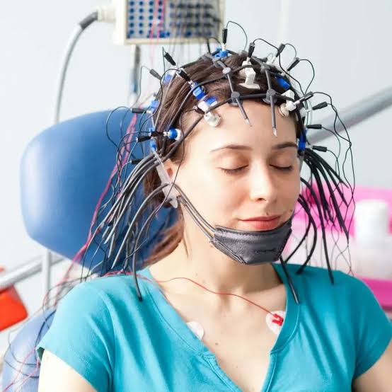
During the test, electrodes placed on different parts of your scalp pick up these signals and send them to the recording equipment. The result appears as a series of brain-wave patterns that can be reviewed by a trained doctor.
EEG therefore gives us information about brain function.
That is different from an MRI or CT scan, which mainly shows brain structure.
This difference is important. A person can have a normal-looking brain scan but still have abnormal electrical activity related to seizures. In other situations, an MRI or CT may reveal the structural cause of symptoms while EEG answers a different question.
When Is an EEG Needed?
Doctors do not order EEG simply because someone has a headache, dizziness, or every type of neurological symptom.
The test is most useful when the history suggests that unusual electrical activity in the brain may be causing the problem.
After a First or Suspected Seizure
A first suspected seizure is one of the most common reasons for an EEG.
Your doctor will want to understand exactly what happened:
- Did you lose awareness?
- Was there shaking or stiffening?
- Did you suddenly stop responding?
- Did someone notice unusual eye or head movements?
- Were you confused afterward?
- Did you bite your tongue or injure yourself?
- What were you doing immediately before the event?
EEG findings can support a diagnosis of epilepsy and provide information about seizure type or epilepsy syndrome.
NICE recommends considering a routine awake EEG when the history and examination suggest an epileptic seizure. If an EEG is requested after a first seizure, it recommends performing it as soon as possible, ideally within 72 hours.
If seizures have already occurred, keeping track of possible seizure triggers such as missed sleep, missed medication, illness, or other individual patterns can also give your neurologist useful context.
For Repeated Blackouts, Staring Spells, or Loss of Awareness
Not every seizure causes dramatic shaking.
Some seizures may cause:
- a brief blank stare;
- sudden loss of awareness;
- unusual repetitive movements;
- unexplained confusion;
- short memory gaps;
- nighttime episodes;
- sudden jerks;
- episodes in which the person does not respond normally.
At the same time, fainting, heart rhythm problems, metabolic problems, panic attacks, and other conditions can sometimes resemble seizures.
This is why distinguishing seizure vs. syncope cannot be based on EEG alone. The description of the episode, examination, medical history, and sometimes heart testing or other investigations are equally important.
When Someone With Epilepsy Needs More Information
EEG may also be recommended after epilepsy has already been diagnosed.
A neurologist may want to:
- classify the seizure type;
- identify where seizures may be starting;
- investigate continuing episodes despite treatment;
- clarify events that do not behave like the patient’s usual seizures;
- obtain a longer recording;
- collect information before selected epilepsy procedures or surgery.
In these situations, a routine EEG may not provide enough information. Ambulatory EEG or video EEG may be more helpful.
After a Stroke or TIA-Like Episode
This part is important because the role of EEG is sometimes misunderstood.
An EEG is not normally the main test used to diagnose an acute stroke or TIA. Brain and vascular imaging such as CT or MRI, together with urgent neurological assessment, are much more important for that question.
EEG may become useful when doctors are asking a different question, such as:
- Did this episode actually represent a seizure rather than a TIA?
- Is someone having seizures after a stroke?
- Is ongoing confusion caused by seizure activity?
- Could non-convulsive seizures explain reduced responsiveness?
Research also shows why clinical judgment matters: EEG abnormalities in people with transient neurological symptoms do not always cleanly distinguish seizure from TIA.
If someone suddenly develops weakness on one side, facial drooping, difficulty speaking, severe imbalance, sudden vision loss, or other possible stroke symptoms, they should seek emergency medical care immediately rather than waiting for an outpatient EEG.
What Happens During an EEG Test?
The equipment can look complicated, especially when several wires are attached to the head. The procedure itself is usually straightforward.
1. Your Scalp Is Measured and Prepared
You will usually sit in a comfortable chair or lie on a bed.
The EEG technologist measures and marks different positions on your scalp so the electrodes can be placed correctly.
2. Electrodes Are Placed on the Scalp
Small electrodes are attached with a special paste, gel, adhesive, or an electrode cap.
Nothing is inserted into your brain.
The electrodes are sensors. They pick up the electrical signals your brain is already producing.
3. Your Brain Activity Is Recorded
You will usually be asked to relax and remain reasonably still.
The technologist may ask you to open or close your eyes at different points.
The machine records the signals detected by the electrodes as brain-wave patterns.
4. You May Be Asked to Breathe Deeply or Look at Flashing Lights
Some routine EEGs include activation procedures.
You may be asked to breathe deeply and regularly for a short period or look at a flashing light.
These steps can bring out brain-wave changes that may not appear while you are simply resting.
For people who are susceptible to seizures, these procedures can occasionally trigger one. The test is performed in a setting where trained staff can respond if this happens.
5. The Electrodes Are Removed
After the recording, the electrodes are removed.
Some paste or adhesive may remain in the hair, so you may want to wash your hair afterward.
Most people can return to their usual activities unless they received a sedative or were given specific instructions by their healthcare team.
Does an EEG Hurt or Send Electricity Into the Brain?
No electricity is sent into your brain during a standard EEG.
This is one of the most common worries patients have.
The electrodes act like microphones for electrical activity: they detect what is already happening rather than creating it.
An EEG is therefore considered a non-invasive and generally painless test. You may notice the sticky paste, pressure while the scalp is prepared, or mild irritation afterward, but the recording itself should not hurt.
How Should You Prepare for an EEG?
Your testing center should give you instructions for your particular EEG.
Common instructions include:
- Wash your hair before the appointment.
- Keep the hair free of oils, gels, sprays, waxes, and styling products.
- Tell your doctor about all medicines and supplements you take.
- Do not stop medicines unless your doctor specifically instructs you to.
- Follow any instructions about caffeine.
- Follow sleep instructions carefully if you are having a sleep-deprived EEG.
Preparation varies depending on the test, so your own neurologist’s or EEG laboratory’s instructions should take priority over general advice online.
What Are the Different Types of EEG?
Not every EEG is the same.
The type your doctor chooses depends on what they are trying to capture.
Routine EEG
This is the standard outpatient EEG.
A routine recording commonly lasts around 20–40 minutes, although the complete appointment is longer because placing and removing the electrodes takes additional time.
It is often the first EEG used when epilepsy or seizures are suspected.
Sleep-Deprived EEG
Some seizure-related abnormalities are easier to detect during drowsiness or sleep.
Your doctor may therefore ask you to sleep less than usual before the EEG.
If your routine EEG is normal but epilepsy is still suspected, guidelines support considering a sleep-deprived EEG.
Ambulatory EEG
An ambulatory EEG records brain activity for much longer while you continue many normal daily activities.
The recording may continue for a day or more.
This gives the test a better opportunity to capture an event that happens too infrequently to appear during a short routine recording.
Video EEG Monitoring
Video EEG records two things at the same time:
- what your brain is doing electrically; and
- what your body is doing during the event.
This can be extremely useful when doctors need to determine whether a repeated episode is an epileptic seizure, identify seizure type, or locate where seizures begin.
Longer video EEG monitoring may take place in an epilepsy monitoring unit when more detailed evaluation is needed.
What Do EEG Results Mean?
A neurologist or another appropriately trained specialist reviews the patterns recorded during the EEG.
The report may describe the recording as normal or may identify findings such as:
- slowing;
- asymmetry;
- spikes or sharp waves;
- epileptiform discharges;
- focal abnormalities;
- generalized abnormalities.
The important point is that an EEG report should never be interpreted in isolation.
Your neurologist needs to compare it with:
- what actually happened during your episode;
- eyewitness descriptions;
- your neurological examination;
- your medical history;
- MRI or CT findings when relevant;
- medications;
- previous neurological problems.
An abnormal EEG may strongly support a diagnosis, but not every abnormality means epilepsy.
Can You Have Epilepsy With a Normal EEG?
Yes.
This is one of the most important facts to understand about EEG testing.
A routine EEG records only a limited period of time. Seizure-related electrical abnormalities do not necessarily appear continuously.
Someone may therefore have epilepsy even though the short recording performed that day looks normal.
NICE specifically advises clinicians not to use EEG to exclude epilepsy. If diagnostic uncertainty remains after a normal routine EEG, a sleep-deprived or ambulatory EEG may be considered.
The reverse is also important:
An abnormal EEG does not automatically prove epilepsy.
The report has to make sense when compared with the person’s actual symptoms and clinical history.
EEG vs MRI or CT Scan: What Is the Difference?
Think of these tests as answering different questions.
EEG | MRI / CT |
Records electrical brain function | Shows brain structure |
Particularly useful for seizures | Useful for stroke, bleeding, tumors, structural abnormalities and many other conditions |
Measures brain activity over time | Creates images of the brain |
May be normal between seizures | May show structural causes associated with seizures |
Does not replace brain imaging | Does not directly replace EEG for seizure-related electrical activity |
For people being evaluated after a seizure, both types of information may sometimes be needed.
For someone with suspected acute stroke or TIA, imaging and urgent clinical assessment generally take priority over EEG.
Is an EEG Safe?
EEG is generally considered very safe.
The electrodes themselves do not cause seizures because they do not stimulate the brain.
However, some EEGs deliberately include flashing lights or deep breathing to see whether these conditions reveal abnormal electrical patterns. In a person already susceptible to seizures, those procedures can occasionally provoke a seizure.
The staff performing the test are trained to recognize and manage this possibility.
Minor temporary scalp irritation from electrode paste or adhesive can also occur.
When Should Symptoms Be Treated as an Emergency?
An outpatient EEG should never delay emergency treatment.
Seek urgent medical help if someone has:
- sudden weakness or numbness affecting one side of the body;
- sudden difficulty speaking or understanding speech;
- sudden facial drooping;
- sudden severe loss of balance or coordination;
- sudden significant vision loss;
- a first major seizure;
- a seizure that lasts unusually long;
- repeated seizures without proper recovery between them;
- prolonged unconsciousness or severe confusion afterward;
- a significant injury during a seizure.
In these situations, the immediate priority is medical stabilization and determining what caused the symptoms. EEG may become part of the evaluation later if appropriate.