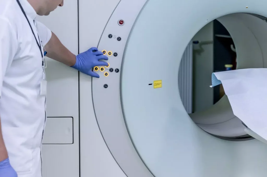
What is an MRI?
Table of Contents
ToggleMRI stands for Magnetic Resonance Imaging. An MRI machine takes very clear pictures of your brain using a magnetic field.
https://www.shutterstock.com/image-photo/mri-machine-hospital-room-482984881
MRI machines are graded based on the strength of the magnetic field they can produce. If an MRI can create a powerful magnetic field, it produces very clear pictures of the Brain. Magnetic fields are measured in units called “Tesla.”
Here are some typical MRI (Magnetic) Strengths:
- 1.5 Tesla – Most common.
- 3 Tesla – Almost all Epilepsy centres have a 3Tesla MRI machine.
- 7 Tesla – Strongest MRI approved for human use so far. Only manufactured by Siemens. Only a few hospitals in the world have this machine.
As you can imagine, a low strength MRI can fail to display tiny Brain abnormalities. Therefore, when a patient comes to an Epilepsy center, frequently, the first thing done is to get a brain MRI at high strength (at least 3Tesla).
If the MRI does not display an abnormality, does it mean that I do not have Epilepsy?
No.
In many cases, the brain abnormality can be so small that it cannot be detected even with an MRI. A 3Tesla MRI (the most common MRI at Epilepsy centres), fails to display brain abnormalities in roughly 50% of patients with Epilepsy.
As MRI strength continues to grow (e.g. 9T & beyond), the number of people with Epilepsy in whom we cannot see the problem will decrease.
If the MRI displays an abnormality, does it mean that I definitely have Epilepsy?
No.
No one is perfect. Any MRI done on a person is bound to show some variations from normal.
https://www.shutterstock.com/image-photo/brain-disease-diagnosis-medical-doctor-diagnosing-1525567082
Beyond a certain age (say 40 years), the effects of ageing become visible on the MRI. There may be changes caused by high blood pressure, by diabetes, by trauma, and so on. Very few of these things seen on an MRI are capable of producing seizures.
The critical question is: Does the thing seen on the MRI look like it can produce seizures? Answer this question requires expertise. A trained radiologist takes a very close look at the MRI and tries to identify it’s exact nature (more below). If he/she thinks that the abnormal thing is capable of producing Epilepsy, he labels it “Epileptogenic”.
If an abnormal thing seen on the MRI is thought to be capable of producing Epilepsy, it is called an “Epileptogenic Lesion”.
What are the different kinds of “Epileptogenic Lesions” seen on an MRI?
Let’s talk about the most common ones. Let’s list them first, before looking at what the MRIs look like:
| Epileptogenic Abnormality | Meaning |
|---|---|
| Lissencephaly | Fewer wrinkles on the brain |
| Polymicrogyria | Too many wrinkles on the brain |
| Focal Cortical Dysplasia | Part (focal) of the brain surface (cortex) is abnormally formed (dysplasia) |
| Nodular Heterotopia | Abnormal bunch of cells stuck in strange locations inside the brain |
| Tuberous Sclerosis | The brain has many large bumps (tubers) on the surface and nodules deep inside (subependymal nodules) |
| Mesial Temporal Sclerosis | The inner surface of the temporal lobes (behind our ears) is scarred |
| Sturge-Weber | There are too many blood vessels over some parts of the brain, and the brain surface below these blood vessels is abnormal |
| Cavernoma | A bunch of thin-walled blood vessels that slowly leak blood |
| Gliosis | Scarring of the brain due to any reason, e.g. trauma due to a vehicular accident, stroke, etc. |
https://upload.wikimedia.org/wikipedia/commons/9/9d/Lissencephaly.jpg
https://upload.wikimedia.org/wikipedia/commons/1/1d/TSC.png
https://www.shutterstock.com/image-photo/neurosurgeon-cutting-brain-specimen-abnormal-vessel-1163324815
https://upload.wikimedia.org/wikipedia/commons/1/1d/TSC.png
Even if an Epileptogenic lesion is not found, can the MRI help in diagnosing my disease?
Yes!
In some people, Epilepsy is caused by a chemical (Metabolic) problem. In these people as well, characteristic MRI changes (e.g. deposition of heavy metals) may help in diagnosing your condition.
For example, see the list below. This is just for reference, and not to be memorized:
| Metabolic Disease Producing Epilepsy | Cause | MRI Findings |
|---|---|---|
| Non-Ketotic Hyperglycinemia | Too much of a substance called Glycine | Swelling and formation of fluid balloons in structures called the pyramidal tract, MCP, and dentate nuclei |
| Maple Syrup Disease | Problems in handling some kinds of proteins |
Swelling of the entire brain The white matter can become extremely bright |
| Zellweger Syndrome | Abnormal bubbles inside cells |
Polymicrogyria & heterotopia (just behind the ears) White matter has not developed the normal coating of fat |
| Menke’s Disease | Accumulation of copper |
Skull bones may be in pieces (wormian bones) Chronic subdural bleeds Brain shrinks White matter swells Copper accumulates in basal ganglia Blood vessels become tortuous |
Note: MRI findings in other metabolic causes of seizures are given below, just for completion. This part is not necessary for you to read:
| Metabolic Disease Producing Epilepsy | Cause | MRI Findings |
|---|---|---|
| Methyl-Malonic Aciduria | Problems in handling some kinds of proteins & fats | Swelling of small structures deep inside the brain called Globus Pallidi |
| Glutaric Aciduria Type 1 | Problems in handling some kinds of proteins | Grooves on the side of the brain (Sylvian fissures) become very wide |
| Hydroxy Glutaric Aciduria | Unknown |
Swelling of white matter just below the brain surface (U-fibers) Deep structures (especially Dentate Nucleus) show injury |
| Molybdenum Cofactor Deficiency | Accumulation of a toxic substance called sulfite | The surface of the brain becomes very bright as if it is starved of oxygen |
| Congenital Creatine Deficiency | Low levels of essential enzyme activity | No creatine inside cells |
| MELAS | Abnormal mitochondria inside cells | Stroke-like lesions that move from one place to another |
| Leigh’s Disease | Unknown | Deep structures start to die (Basal Ganglia & Dentate nucleus) with accumulation of a chemical called lactate |
| Neuronal Ceroid Lipofuscinosis | Accumulation of fatty pigments (lipofuscin) |
The back of the brain shrinks White matter can become “dark” |
Caution: This information is not a substitute for professional care. Do not change your medications/treatment without your doctor’s permission.
